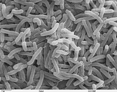
Vibrio Cholerae is the bacteria responsible for cholera cases all around the
world. The bacterium is said to be originated from the Bengal region in India.
So far, two serotypes of Vibrio cholerae,
O1 & O 139 have been identified as the cause of cholera ourbreaks. El Tor
is a new strain of Vibrio cholerae that emerged recently.
People who is infected by El Tor are usually asymtomatic or only experience mild
cholera symptoms.
Where is Vibrio cholerae found?
The cholera bacterium is usually found
in water or food sources that have been contaminated by faeces from a person or
animal infected by cholera. Cholera is most likely to be found and spread in
places with inadequate water treatment, poor sanitation and inadequate hygiene.
The cholera bacterium may also live in
the environment in brackish rivers and
coastal waters (especially in the area where fresh water meets the sea).
Shellfish may also transmit cholera bacterium.
In the past few years, cholera has been
a minor threat to developed countries such as USA and European countries mainly
because of advanced water treatment system and sanitation.
 |
 |


Mechanism of Vibrio cholerae
Most bacteria,
when consumed, do not survive the acidic conditions of the human stomach. The
few surviving bacteria conserve their energy and stored nutrients during the
passage through the stomach by shutting down much protein production. When the
surviving bacteria exit the stomach and reach the small intestine, they need to
propel themselves through the thick mucus that lines the small intestine to get
to the intestinal walls, where they can thrive. V. cholerae bacteria start up
production of the hollow cylindrical protein flagellin to make flagella, the
cork-screw helical fibers they rotate to propel themselves through the mucus of
the small intestine.
Once the cholera
bacteria reach the intestinal wall, they no longer need the flagella to move.
The bacteria stop producing the protein flagellin, thus again conserving energy
and nutrients by changing the mix of proteins which they manufacture in
response to the changed chemical surroundings. On reaching the intestinal wall,
V. cholerae start producing the toxic proteins that give the infected person a
watery diarrhea. This carries the multiplying new generations of V. cholerae
bacteria out into the drinking water of the next host if proper sanitation
measures are not in place.
Microbiologists
have studied the genetic mechanisms by which the V. cholerae bacteria turn off
the production of some proteins and turn on the production of other proteins as
they respond to the series of chemical environments they encounter, passing
through the stomach, through the mucous layer of the small intestine, and on to
the intestinal wall. Of particular interest have been the genetic mechanisms by
which cholera bacteria turn on the protein production of the toxins that
interact with host cell mechanisms to pump chloride ions into the small
intestine, creating an ionic pressure which prevents sodium ions from entering
the cell. The chloride and sodium ions create a salt-water environment in the
small intestines, which through osmosis can pull up to six litres of water per
day through the intestinal cells, creating the massive amounts of diarrhea. The
host can become rapidly dehydrated if an appropriate mixture of dilute salt
water and sugar is not taken to replace the blood's water and salts lost
in the diarrhea.
By inserting
separately, successive sections of V. cholerae DNA into the DNA of other
bacteria, such as E. coli that would not naturally produce the protein toxins,
researchers have investigated the mechanisms by which V. cholerae responds to
the changing chemical environments of the stomach, mucous layers, and
intestinal wall. Researchers have discovered a complex cascade of regulatory
proteins controls expression of V. cholerae virulence determinants. In
responding to the chemical environment at the intestinal wall, the V. cholerae
bacteria produce the TcpP/TcpH proteins, which, together with the ToxR/ToxS
proteins, activate the expression of the ToxT regulatory protein. ToxT then
directly activates expression of virulence genes that produce the toxins,
causing diarrhea in the infected person and allowing the bacteria to colonize
the intestine. Current research aims at discovering "the signal that makes
the cholera bacteria stop swimming and start to colonize (that is, adhere to
the cells of) the small intestine."
Genetic Structure of Vibrio
cholerae
Amplified fragment
length polymorphism fingerprinting of the pandemic isolates of V. cholerae has
revealed variation in the genetic structure. Two clusters have been identified:
Cluster I and Cluster II. For the most part, Cluster I consists of strains from
the 1960s and 1970s, while Cluster II largely contains strains from the 1980s
and 1990s, based on the change in the clone structure. This grouping of strains
is best seen in the strains from the African continent.
Which language does the 'vibrio cholerae' derived from? Can i know the meaning of 'vibrio'?
ReplyDeleteHello there. Thanks for your time for commenting our blog.
ReplyDeleteWell, actually V. cholerae was first isolated as the cause of cholera by Italian anatomist Filippo Pacini in 1854, but his discovery was not widely known until Robert Koch, working independently 30 years later, publicized the knowledge and the means of fighting the disease. So, it is believed that the language of the term "Vibrio cholerae" is derived from Latin.
"Vibrio" has the meaning of:
a genus of gram-negative, short, motile, curved or straight rods in the family Vibrionaceae of bacteria. The word "vibrio" in Latin means "to quiver."
Hope these information do help you ! :D
I'm confused ! This bacteria was originated from the bengal region of India or bangladesh? I remember reading the wiki about this and the source said that it was first identified in bangladesh although recently the infection caused by this bacteria have become rare and limited to parts of Bangladesh and india
ReplyDeleteWe have actually different sources of info that determines the source of cholera is from Bengal as well as Bangladesh. The truth is we are also not so sure about the whereabouts of the origin.But since Bengal and Bangladesh are quite near to each other, cholera is most likely to have started between or around these areas.
Delete